
Shoulder Arthroscopy Surgery
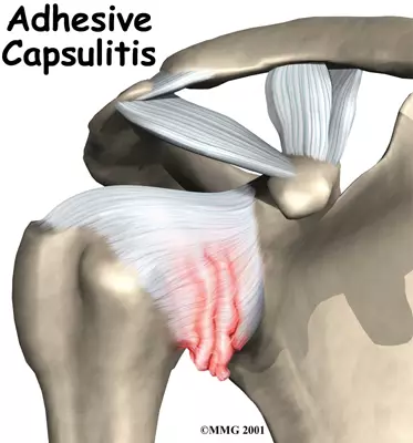
A painful shoulder can make even simple tasks difficult to do. You might find it hard to use your arm or your hand when you have pain. But getting the right treatment can help. Shoulder pain is any pain in or around your shoulder joint. You may feel the pain most when you reach behind your back or overhead. There are many reasons why you may get a painful shoulder.


")
")
")

Complex Trauma Injury
Our experts provide advanced care for complex trauma injuries, ensuring effective treatment and recovery for severe and multifaceted fractures and wounds.
View Details
Arthroscopy & Sports Injuries
We specialize in minimally invasive arthroscopy and the treatment of sports injuries, helping athletes return to peak performance quickly and safely.
View Details
Joint Preservation
Our joint preservation techniques focus on maintaining natural joint function and delaying the need for joint replacement through innovative treatments.
View Details
Back Pain Management
Our skilled surgeons perform advanced spine surgeries to treat various spinal disorders, providing relief and improved spinal health.
View Details